Friday, April 20, 2007

Copy Catsprinkles Red Velvet Cupcake Recipe

Number of figures

On many occasions we need to make a huge amount of data to a JTable with a scroll very slow and consume large amounts of memory and resources.

Then the following realia manual that explains how this task in the most optimal possible. This manual is essential to properly utiizar a large volume of data in an application in java JTable

Wednesday, April 11, 2007

Could Ipod Earphones Be Used As Ps3 Mic

Cytoskeletal Molecular Motors

The Cytoskeleton

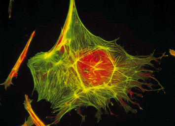
In ordinary preparations of visible light microscopy or electronic, the cytoskeleton appears transparent and therefore invisible. Generally not as drawn in the schemes of the cell but is an important cellular component, complex and dynamic. The cytoskeleton maintains the shape of the cell, "anchor" the organelles in place and move part of the cell processes of growth and mobility.

There

various types of protein filaments that constitute the cytoskeleton: microfilaments, microtubules and intermediate filaments.

The microtubules are composed of subunits of a protein called tubulin and are often used by the cell to maintain its shape, are also the largest component of cilia and flagella.

The microfilaments are composed of subunits of the protein actin. They are about one third the diameter of the microtubule and are often used by the cell to change its structure so as to maintain it.

A large number of proteins associated with the cytoskeleton that control its structure both through the guidance and direction of the filaments and groups of their movement. A particularly interesting group of proteins associated with the cytoskeleton are "other" phones, such as myosin (an "engine" that moves actin filaments) and kinesin (a "motor" for microtubule).


The three components cytoskeleton are interconnected and form a lattice, which extends from the cell surface to the core. This system is built on a common architectural model is in an amazing variety of natural systems and is known as tensional integrity (tensegrity). This expression indicates that the system stabilizes itself mechanically, because of how the compression and tension forces are distributed and balanced within the structure.


structures that respond to this model of tensional integrity not reach mechanical stability and resistance of individual members but by the way the whole structure distributes and balances mechanical stresses. In these structures the tension is transmitted seamlessly through all elements estructurales.En other words, an increase of stress in any element of the structure is felt in all others. This overall increase in pressure is balanced by an increase in the compression of certain elements all over the structure. An archetypal example of these structures are geodesic domes Buckminster Fuller.

should be noted that the universal rules governing the construction applies to the formation of organizational structures, from molecules to tissues.

Centrioles and Basal Bodies.

are basically the same thing, are interconvertible structures. A centriole is made up of nine bundles of microtubules triplets. A triplet contains a complete microtubule fused to two incomplete. The basal bodies are related with cilia and flagella where, as centrioles, are associated with the cytoskeleton.

Centrioles play a vital role in cell division. They are surrounded by a dense material that stains the pericentriolar material , from which microtubules originate, even in cells without centrioles microtubules originating from this material.

Centrioles are often known for their role in cell division. They are members of the centrosome consisting of two perpendicular centrioles. The centrioles appear to determine the position of material pericentriolar, which in turn affects cell polarity. Note that the functional centrosome is typically from an embryo sperm fertilizing.

To expand on this topic see this literature.


Cytoskeleton Tutorial

Microtubules, microfilaments and intermediate filaments


The cytoskeleton

http://www.biologia.arizona.edu/cEll/tutor/cyto/page1.html

Cytoskeleton

http://www2.uah.es/biologia_celular/LaCelula/Cel5CK.html

CancerQuest

http://cancerquest.org/index.cfm?page=46 & ; lang = English & English CHANGETABLE =

Cell Biology Manual

http://w3.cnice.mec.es/eos/MaterialesEducativos/mem2001/biologia/citoplasma/esqueleto.htm

interaction between actin and other molecules, such as myosin -


Did you know ...?

actin filaments play an important role in cell motility . Among its properties emphasizes their polarity, which is behavior different from its two extremes: while one is biased or lengthens (positive end), the other tends to shorten or depolymerize (negative end). In addition, actin filaments are involved in phagocytosis in the process of muscle contraction and production of cytoplasmic flows .

For their part, intermediate filaments are highly resistant protein fibers that play a structural role in cells, especially those who are subjected to significant mechanical stresses. Depending on the cell type predominantly one or the other (neurofilament in cells nerve, vimentin in blood vessels, etc.) It is this diversity is of immense use in source characterization in cancer metastasis, thus, determining the type of intermediate filament we know the tissue where they produced the original tumor . But then, in addition, these filaments may help in the prenatal diagnosis congenital malformations.

Que Piercing Tiene Andy Sixx


DINAIN, kinesin and Dynamin

The operation of actin-myosin complex becomes clear that cytoskeletal structures are ideal as support points, guides and transmission lines of the forces developed by molecular motors in cells. Not surprisingly, therefore, that the microtubules more rigid and stronger than actin filaments, constitute support for the operation of other molecular motors. And in retrospect, does not cause much astonishment that such engines are designed under the same principle as myosin. In fact, at least in the first case we will discuss below, are very similar (Table 1).


The DINAIN - protein force - is another mecanoenzima that uses energy from ATP hydrolysis to temporarily change its shape. It is also a large molecule composed of several subunits, the largest of which are two globular heads with a weight of 410 000 daltons each, in which resides the ATPase activity. This activity increased substantially in the presence of microtubules. The DINAIN was initially identified as the engine driving the movement of vibrating appendages called cilia and flagella that certain cells have. More recently it became obvious that there are other ways DINAIN distributed in the cytoplasm of a wide variety organisms.

The hallmark of DINAIN is its ability to move on microtubules in the same way as does myosin on actin filaments (Figure V.4). It is worth mentioning that this interaction is on the outside and not by the light of microtubules close as anyone could reasonably think. It appears that DINAIN heads are secured to a site on the wall of microtubules, then suddenly change shape, and then off to straighten and lock in a new site, constantly repeating the cycle while ATP available.


The ATP-ase in the Exonemo breaks the molecules of ATP and thus obtains energy for the movement of sperm. There is a theory by which the sperm move through a mechanism oscillator. Alternating sweeps right and left, the microtubules of the tail.


Spermatozoa.


sperm metabolism is aerobic and Cl and Mg ions promote motility. While the Ca +2 ion inhibits motility.

For proper motility is necessary to open Ca +2 channels and there is a change in the concentration of nucleotides in the Exonemo in the proximal portion of the tail.
In addition, any substance that inhibits the enzyme activity in sperm is a toxic substance for which hinders their motility.

The movement begins in the proximal portion of the tail (axoneme) and goes into the final. The sheath protein controls the tail beating, so it is not very sharp. There are a number of glands that release fundmental seminal fluid for sperm travel. The glands are in the path of it. So will acquire motility along the epididymis, the vas deferens, with the secretions of various glands.

In the bull, sperm speed is 72 microns per second and man, 35 to 50. The optimum pH is 7.5.

The fetal testis is an endocrine organ, acting as such. Is very different from ovarian feta, because the testicle produces a large amount of hormones. Among them:
• Testosterone: synthesized by fetal Leydig cells.
• AMH (AMH), synthesized by Sertoli cells.

sperm structure

The sperm will have a head, which houses the core and a structure that allows motility (tail). In the anterior nucleus is the acrosome, which is under the plasma membrane and leads enzymes released at fertilization. Behind the head, the neck is the region containing the centriole.


Cola: In the thickest part is the axoneme, where mitochondria. After the tail is tapered. Along the line, there are common structures, microtubules. There are 9 pairs of peripheral microtubules and a central pair. They are composed of protofilaments. One of the rings are 13 and 11 protofilaments.
The protofilaments consist of:
• Tubulin : two dimers (a1, a2 [also a, b])
• Histone .
• dynein : Is an ATPase. The role is to act on the mitochondria and ATP provide the sperm movement.


dynein arms are very important. Kartagener syndrome in man (also been studied in rats) individuals are unable to synthesize dynein. Its non-motile sperm, as well the hair cells of the respiratory tract and have great rates of respiratory diseases. Their hearts are on the right side "situs inversus."

References. E. Frixione

Meza I. (2006) Living Machines How Cells are moved? Fondo de Cultura Economica . Mexico, DF ISBN 6-4988-5 968-l

Electronic References.

Reproduction.

http://web.educastur.princast.es/proyectos/biogeo_ov/2BCH/B4_INFORMACION/T409_REPRODUCCION/informacion.htm

floating fertilization.

http://ciencia.nasa.gov/headlines/images/fertility/graveffects_med_sp.gif

New scientific.

http://www.novaciencia.com/2006/11/28/pildora-para-bloquear-la-eyaculacion/

Medline Plus

http://www.nlm.nih. gov/medlineplus/spanish/ency/esp_imagepages/9875.htm

Recuperados on 10 April 2007.